Do male and female brains really differ?

Research uncovers the truth about brain differences between men and women, cutting through historical bias and gender politics.
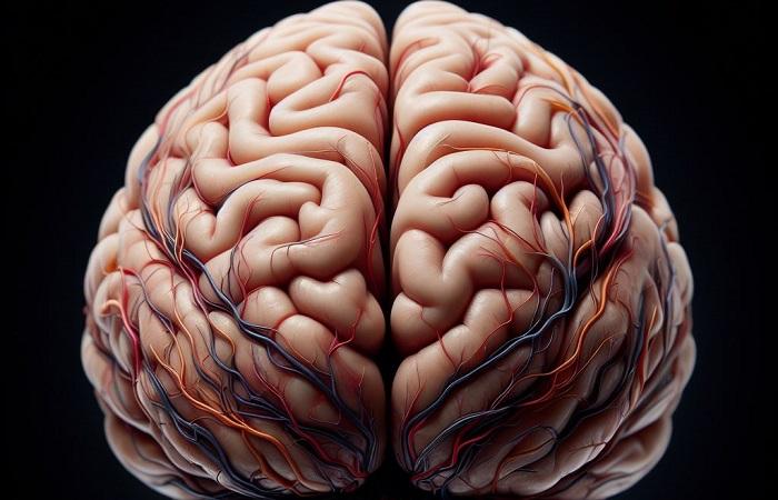
27740fcc e347 4349 a023 32ffd140ae97

The question of whether there are differences between the brains of men and women has been debated for centuries and has implications for various fields, such as psychology, education, and medicine.

However, the answer is not simple or clear-cut, as the brain is a complex and dynamic organ, that is influenced by many factors, such as genes, hormones, environment, and experience. Moreover, the brain is composed of billions of cells, or neurons, that communicate with each other through electrical and chemical signals, and form networks and circuits that underlie various functions and behaviors.

The facts

One of the most obvious differences between the brain cells of men and women is their genetic makeup. On average, males and females showed greater volume in different areas of the cortex, the outer brain layer that controls thinking and voluntary movements.

Females have greater volume in the prefrontal cortex, orbitofrontal cortex, superior temporal cortex, lateral parietal cortex, and insula. These regions are involved in various cognitive and emotional processes, such as decision-making, social behaviour, language, and memory.

Males have greater volume in the occipital cortex, the posterior parietal cortex, and the amygdala. These regions are involved in visual perception, spatial processing, and emotional reactions.

However, these differences in brain volume do not necessarily translate into differences in brain performance, as other factors, such as connectivity, plasticity, and efficiency, may also play a role. Moreover, these differences are not absolute, as there is a lot of overlap and variation among individuals of the same and different sexes.

The arguments

Another way to compare the brain cells of men and women is to look at their function, or how they respond to various stimuli and tasks. One method to measure this is to use brain imaging techniques, such as functional magnetic resonance imaging (fMRI), which can detect changes in blood flow and oxygen levels in different brain regions, indicating their level of activity.

Several studies have used fMRI to examine the brain activity of men and women while they perform various cognitive and emotional tasks, such as verbal, spatial, memory, and face recognition tasks. Some of these studies have found sex differences in the patterns of brain activation, suggesting that men and women may use different strategies or recruit different brain regions to solve the same problems.

For example, one study found that women showed more activity in the left inferior frontal gyrus, a region involved in language processing, while men showed more activity in the left hippocampus, a region involved in memory formation when they performed a verbal fluency task.

Another study found that women showed more activity in the right fusiform gyrus, a region involved in face perception, while men showed more activity in the right inferior parietal lobule, a region involved in spatial attention when they performed a face recognition task.

However, these differences in brain activation do not necessarily imply differences in brain ability, as the same outcome can be achieved by different neural pathways, and the same neural pathway can be used for different outcomes.

Moreover, these differences are not consistent, as they may depend on the type, difficulty, and context of the task, as well as the individual characteristics and preferences of the participants.

A third way to compare the brain cells of men and women is to look at their activity, or how they communicate with each other and form networks and circuits. One method to measure this is to use electroencephalography (EEG), which can record the electrical activity of the brain cells, or magnetoencephalography (MEG), which can record the magnetic fields generated by the brain cells.

These techniques can capture the frequency, amplitude, and synchronization of the brain waves, which reflect the level, intensity, and coordination of the brain cell activity.

For example, one study found that women had higher alpha power, a measure of brain relaxation, than men, while men had higher beta power, a measure of brain alertness, than women, when they were at rest.

More from Qonversations

TalkingPoint

Gene editing

CRISPR-Cas9 and Gene Editing: Are we redefining nature’s blueprint?

TalkingPoint

Screenshot 2024 12 04 at 1.58.18 PM

Digital ghosts: The controversial rise of AI resurrections in Mexico

TalkingPoint

Trump and Femi

Are conservatives really happier? New study explores the politics of happiness and psychological richness

TalkingPoint

Global warming red

Is humanity ignoring the warning signs of climate catastrophe?

Front of mind